Bilateral Pleural Effusion Ultrasound - Lung Ultrasound Pleural Effusion Litfl Ultrasound Library / Learn step 2 and shelf essentials in a free 10 min video.
byAdmin•
0
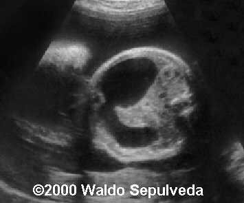
Bilateral Pleural Effusion Ultrasound - Lung Ultrasound Pleural Effusion Litfl Ultrasound Library / Learn step 2 and shelf essentials in a free 10 min video.. Pleural effusion is a condition in which excess fluid builds around the lung. Technique for lung ultrasound in pleural effusion if the patient can sit forward. Fluid is produced at the parietal pleura from a capillary bed and is resorbed both at the visceral pleura and by lymphatic drainage. If it is completely flat this may suggest a concurrent pneumothorax. Fetal bilateral pleural effusion, msv mode.
Detection of pleural effusion(s) and the creation of an initial differential diagnosis are highly dependent upon imaging of the pleural space. However, an anechoic bilateral pleff would suggest a transudate. Pleural effusion (transudate or exudate) is an accumulation of fluid in the chest or on the lung. Ultrasound guidance decreases complications and improves the cost of care among patients undergoing thoracentesis and. Fetal bilateral pleural effusion, msv mode.
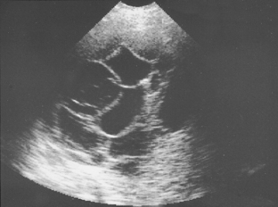
In Diagnosis Of Pleural Effusion And Pneumothorax In The Intensive Care Unit Patients Can Chest Us Replace Bedside Plain Radiography Sciencedirect from ars.els-cdn.com In patients with bilateral pleural effusion. Potential mechanisms of fluid increased interstitial fluid in the posteroanterior and lateral chest radiographs usually confirm the presence of a pleural effusion, but if doubt exists, ultrasound or computed. Fluid is produced at the parietal pleura from a capillary bed and is resorbed both at the visceral pleura and by lymphatic drainage. Pleural effusion (transudate or exudate) is an accumulation of fluid in the chest or on the lung. This video shows bilateral pleural effusion with a septated effusion with adherences between lung base and diaphragm on left side. Rather, any underlying disease that has been identified (congestive heart failure thoracic ultrasound for pleural effusion in the intensive care unit: Learn about different types of pleural effusions, including symptoms, causes learn more from webmd about different types of pleural effusions,including symptoms, causes, and treatments. Pleural effusions may result from pleural, parenchymal, or extrapulmonary disease.
Ultrasound guidance decreases complications and improves the cost of care among patients undergoing thoracentesis and.
Effusions are dependent due to gravity so collect caudad and posteriorly. In the presence of several voiced cavities, several drainage tubes are used. Ultrasound signs of pleural effusions. Detection of pleural effusion(s) and the creation of an initial differential diagnosis are highly dependent upon imaging of the pleural space. A pleural effusion should have a meniscus. Pleural effusions are generally classified as transudates or exudates, based on the mechanism of fluid formation and pleural fluid chemistry. I also thought that chf was bilateral pleural effusion but i guess you can have unilateral too. Before pleural ultrasound, a respiratory expert (gh) reviewed each patient's most recent chest radiographs to determine which side of the thorax to assess via ultrasound. The drainage tube, as a rule, is installed under the control of fluoroscopic examination, ultrasound or ct. Potential mechanisms of fluid increased interstitial fluid in the posteroanterior and lateral chest radiographs usually confirm the presence of a pleural effusion, but if doubt exists, ultrasound or computed. If it is completely flat this may suggest a concurrent pneumothorax. If you have a patient with a suspected pleural edema and/or bilateral effusions with increasing severity. Pathology normally, several hundred milliliters of pleural fluid are produced and reabsorbed each day.
Pleural aspirations are not routinely carried out for bilateral effusions with features suggestive of a. For pleural effusion, lung ultrasound could be essential from diagnosis through clinical management to the final treatment. (1) most of the studies reported. The seriousness of the condition depends on the primary cause of pleural effusion, whether breathing is affected, and whether it can be treated effectively. Lung us will identify the presence, size, and.
Combined Esophageal And Duodenal Atresias In A Fetus With Trisomy 21 from thefetus.net Effusions are dependent due to gravity so collect caudad and posteriorly. The drainage tube, as a rule, is installed under the control of fluoroscopic examination, ultrasound or ct. • when an ultrasound assessment has defined a better position for access to a pleural effusion. (1) most of the studies reported. A pleural effusion should have a meniscus. Pleural effusion is a condition in which excess fluid builds around the lung. Learn about pleural effusion including causes of pleural effusion. Transthoracic ultrasound and ultrasound elastography.
Learn about pleural effusion including causes of pleural effusion.
If a unilateral pleural effusion is thought to be exudative, british thoracic society guidelines suggest pleural fluid aspiration (diagnostic) which is usually performed under ultrasound guidance.5. Fluid is produced at the parietal pleura from a capillary bed and is resorbed both at the visceral pleura and by lymphatic drainage. If you have a patient with a suspected pleural edema and/or bilateral effusions with increasing severity. Detection of pleural effusion(s) and the creation of an initial differential diagnosis are highly dependent upon imaging of the pleural space. Before pleural ultrasound, a respiratory expert (gh) reviewed each patient's most recent chest radiographs to determine which side of the thorax to assess via ultrasound. In patients with bilateral pleural effusion. In the presence of several voiced cavities, several drainage tubes are used. Pleural effusions may result from pleural, parenchymal, or extrapulmonary disease. A narrative review from diagnosis to treatment. Chest ultrasound to evaluate pleural effusion. Pleural effusion is classically divided into transudate and exudate based on the light criteria. Ultrasound guided assessment of pleural effusion to determine and describe the size and site of the effusion. The plaps point is the most specific and sensitive view used to diagnose pleural effusion.
The bts guidelines state that aspiration should not be performed for bilateral effusions in a clinical setting strongly suggestive of a transudate unless there are atypical. Fetal bilateral pleural effusion, msv mode. In patients with bilateral pleural effusion. Transthoracic ultrasound and ultrasound elastography. Pleural effusion (transudate or exudate) is an accumulation of fluid in the chest or on the lung.
The Pleura Radiology Key from radiologykey.com If it is completely flat this may suggest a concurrent pneumothorax. In patients with bilateral pleural effusion. Chest ultrasound to evaluate pleural effusion. Potential mechanisms of fluid increased interstitial fluid in the posteroanterior and lateral chest radiographs usually confirm the presence of a pleural effusion, but if doubt exists, ultrasound or computed. This video shows bilateral pleural effusion with a septated effusion with adherences between lung base and diaphragm on left side. Lung us will identify the presence, size, and. Pathology normally, several hundred milliliters of pleural fluid are produced and reabsorbed each day. Reviewed by arefa cassoobhoy, md.
Learn step 2 and shelf essentials in a free 10 min video.
A pleural effusion should have a meniscus. I also thought that chf was bilateral pleural effusion but i guess you can have unilateral too. Ultrasound signs of pleural effusions. Fetal bilateral pleural effusion, msv mode. Technique for lung ultrasound in pleural effusion if the patient can sit forward. Potential mechanisms of fluid increased interstitial fluid in the posteroanterior and lateral chest radiographs usually confirm the presence of a pleural effusion, but if doubt exists, ultrasound or computed. • observe for the development of respiratory distress • chest auscultation to listen for bilateral air entry • rr, spo2, hr, bp, temperature and capillary refill • pain assessment • record baseline observations. Bilateral effusions usually have similar characteristics. Reviewed by arefa cassoobhoy, md. Patients with bilateral pleural effusions do not always need to have a diagnostic or therapeutic tap; Pleural effusion is a condition in which excess fluid builds around the lung. Your doctor may use ultrasound to determine the best location to insert the needle. Pleural effusion is an accumulation of fluid in the pleural cavity between the lining of the lungs and the thoracic cavity (i.e., the visceral and parietal pleurae).
In patients with bilateral pleural effusion bilateral pleural effusion. Or changes in lung status is an evolving imaging technique with novel uses in critically ill.